- 2 MRT 1,5T und 3T incl. newest Postprocessing-Software
- 2 Multislice CT 16 and 40 Slices incl. central Image-Prozessing via IntelliSpace-Server
- 3 Flat-Panel-Modalities for conventional Radiology
- 1 Angiography-Unit
- 1 Fluoroscopy-Unit
- 1 Mammography-Unit
- 1 Mamma-Stereotaxy-Unit for clarification of breast cancer
- 1 DEXA-Unit for exploration of bone-density
Department of Radiology
The whole range of x-ray examinations and minimal invasive x-ray guided procedures
Diagnostic Modalities
Interventionally Focuses und „Specialities“
- Aortic Stents in cooperation with vascular surgery
- Stents
- Angioplasty
- Embolisations of tumoral- or posttraumatic bleedings
- Thrombektomies und medicamentous lysis for apoplectic Stroke-Patients
- Coiling and Embolisation of aneurysms and haemangiomas of cerebral vessels
- Chemoperfusions e.g. of the liver
- CT-guided drainages, pain-treatment, biopsies and microwave-tumorablations
- CT-guides vertebroplasties
- Mamma-Screening-Clarification with stereotactical aspiration biopsy
Digital flat detection technics to conventional X-ray diagnostics
Thoracic, abdominal and skeletal diagnostic work-up, tomography scans, mammography, radioscopy of regions such as the colon, gastro-intestinal series, Sellink examinations, defecography and micturating cystourography using the most advanced, low-dosage, completely digital flat-panel detector systems.
In cooperation with the Breast Center, the department carries out preoperative labeling of breast cancer tumors, and tumor resection using aspiration biopsy.
Computed tomography
Two multislice CT scanners are used to examine the whole body. The department also conducts a large range of procedures with precisely controlled punctures of tumors and metastases in both soft and boney tissues, along with the puncture of abscesses and the placement of abscess drainage catheters. Furthermore, it uses thermofrequency ablation, principally for the CT-controlled treatment of liver metastases. Vertebroplasty is performed to stabilize osteoporotic sintered vertebrae.
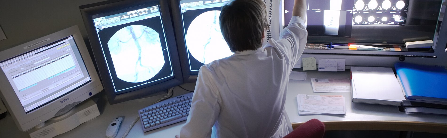
Angiography
The department uses DSA in the completely digital flat-panel detector angiography to examine the arteries and veins of all regions of the body. In addition, it carries out procedures such as dilatation and stent insertion in cases of vascular stenosis, embolization of blood vessels supplying tumors and aspiration of fresh thrombotic material. Aortic stent placement is performed intraoperatively in cooperation with the Department for Vascular Surgery. The stenting of tumor-related (largely venous) vascular stenoses also offers clear benefits to patients.
Magnetic resonance imaging (MRI)
1.5 T and 3 T magnetic resonance imaging scanners can be used for all standard examinations of all regions of the body. The devices combine the latest software and hardware to allow rapid dynamic cardiac MRI. Multimodal imaging, in particular in the region of the bile ducts including MRCP, is also one of the examinations it routinely conducts. MR-supported breast cancer tumor labeling is available in cooperation with the Breast Center. The modern 3T technology also enables the department to perform MR spectroscopy.
The department is also involved in a large number of cooperative projects, some of which are carried out on-site, and some via teleradiology.