The hip osteoarthritis refers to the wear-induced disease of the hip joint. Although osteoarthritis can affect each joint in the body, but the knee and hip are frequently affected as they bear a large part of the body weight.
Etiology
In the case of the hip joint, the cartilage layer which covers the bone of the femoral head of the thigh and the acetabulum of the pelvis prevents friction of both bones. However, misalignments, incorrect loads or injuries can affect this cartilage layer. In the advanced stage, this layer is completely damaged which leads to rubbing of both bones with each other. The result is a limited mobility to a stiff hip and considerable pain.
The wear of the cartilage begins not at an advanced age. In general, it is assumed that the wear and tear of the joints starts from the age of 35.
Investigations
A simple X-ray image is enough normally to confirm the diagnosis "Osteoarthritis". In some cases, an additional MRI is required in order to confirm the diagnosis.
Therapy
Advanced Osteoarthritis is not curable. Through physical therapy, we can initially reduce pain, Improve joint movements and with medications we can control pain and reduces the inflammatory process.
Incorrect and disorganized movement patterns can also be resolved by physical training. Depending on the severity of osteoarthritis, however, intervention is sometimes necessary.
The following 3 chapters at the end of the page explain the different types of Hip replacement.
At Hospital Rummelsberg Prof. Drescher has successfully established a unique therapeutic plan in Germany. Detailed information can be found here.
Hip
Osteoarthritis of the hip Joint
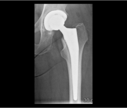
In severe cartilage abrasion, i.e. Joint wear (osteoarthritis, s. X-ray image below) is to discuss together with the patient the joint replacement as a surgical solution. In cases with good bone quality, the cementless joint replacement has been found to be long lasting. We implant the artificial hip joint in a minimally invasive surgical technique-meticulous so that muscles and tendons are not damaged which is optimal for rehabilitation with the best possible results.
By prior planning on computer, we can determine the proper implant size and the optimal position of implantation, so the natural biomechanics is restored which allows quickly achievement of a symmetrical and coordinated gait pattern.
The cementless fixation is achieved by pressing the coated implant „press-fit" in the bone which will be with bone growth completely incorporated in the bone.
With this technique, most of our patients are allowed for full weight after surgery. Here, we use implants have proven over several decades the longest survival rate.
The quality and expertise of our joint surgery allowed our hospital to be certified as one of the first Arthroplasty centers of maximum care in Germany.
In cases of severe cartilage damage, i.e. Osteoarthritis, joint replacement is to be discussed with the patient. In young patients, the cause is often secondary to hip dysplasia or avascular necrosis of the femoral head.
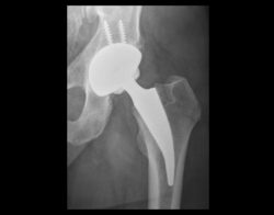
In younger patients, it is very important to preserve as much as possible the bone substance. For this aim , the short stem prosthesis has been developed. The short stem prosthesis which we use in our hospital helps us to preserve most of the neck of the femur which allows better anchor of the prosthesis in bone.
In addition, the short stem is very well suited for the practice of our minimally invasive technology-sparing surgery. Muscles and tendons are not damaged and preserved for a good function. By prior planning on a computer will ensure that the proper implant size and position to be implanted, so the natural biomechanics is recovered. This allows the patient quickly again achieve a symmetrical and coordinated gait pattern. The cementless fixation is achieved by pressing the coated implant „press-fit" in the bone which will be with bone growth completely incorporated.
With this technique, most of our patients are allowed for full weight after surgery.
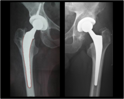
A well-functioning prosthesis needs a firm fixation in the bone of the thigh and pelvis. In the early days of hip Arthroplasty, the cemented anchoring was the standard method, and has achieved good long-term results. Over time the use of the cementless Prosthesis is more frequent implanted. In this case, the fixation mechanism of the prosthesis is achieved by a direct clamping of the prosthesis in the bone.
Due to the long life expectancy of our society, however, many patients have a weak bone quality, as is the case for example in osteoporosis, the bone stock cannot offer enough stability for cementless Fixation der prosthesis. In these cases, the attachment of the prosthesis can be achieved with bone cement; these penetrate a few millimeters into the bone, thereby increasing the area of contact between prosthesis and bone. Thus, the cemented hip prosthesis is in our clinic is still a proven way to allow patients with reduced bone quality a supply of a hip prosthesis.
On the left: cemented (marked in red), on the right: uncemented


